Jryn Eye Clinic Busan (Seomyeon) · SMILE LASIK · EVO+ Visian ICL · Cataract & Retina Center · Tel: +82-51-710-1004
en-US

1:1 Consultation
CLINICAL ARTICLE & GUIDE
Understanding Vitreoretinal Surgery: Procedures & Recovery
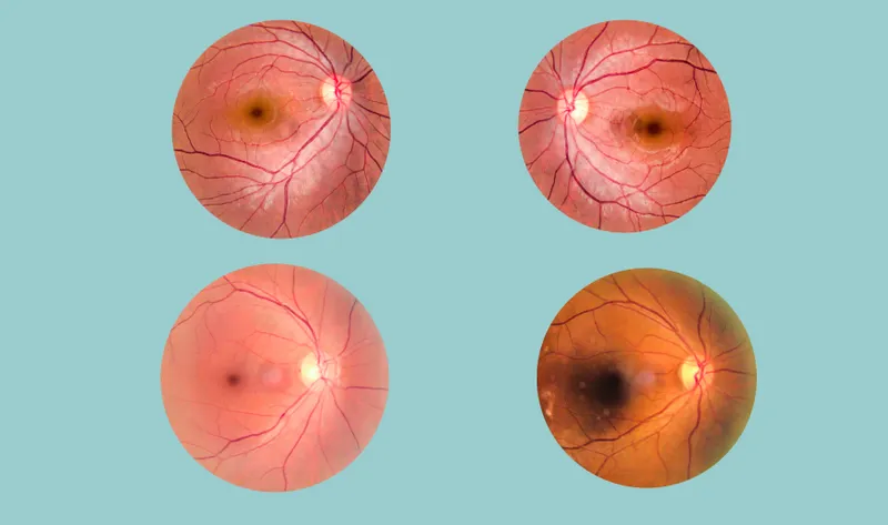
Introduction The human eye is a delicate organ, and for many years, issues deep within the eye were difficult to treat. Today, vitreoretinal surgery has transformed from a high-risk gamble into a precise, vision-saving marvel.
Introduction
The human eye is a delicate organ, and for many years, issues deep within the eye were difficult to treat. Today, vitreoretinal surgery has transformed from a high-risk gamble into a precise, vision-saving marvel. This specialized field of ophthalmology focuses on the structures at the back of the eye, specifically the vitreous and the retina.
The goal of this guide is to help patients understand what happens during these procedures. Whether you are facing an emergency or a scheduled surgery, knowing what to expect during the operation and the recovery period can significantly reduce anxiety and improve your results.

The Evolution of Retinal Care
In the past, retinal diseases often led to permanent blindness. However, modern vitreoretinal surgery (VRS) has evolved into a minimally invasive standard of care. Surgeons now use tools thinner than a human hair to perform complex repairs.
This guide will walk you through the entire journey from the initial diagnosis and the technical steps of the surgery to the essential recovery phase and the global options for affordable, high-quality care.
What is Vitreoretinal Surgery?
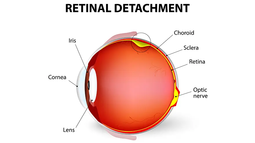
To understand the surgery, you first need to understand the anatomy of the eye.
Anatomy Overview
The Vitreous: This is a clear, gel-like substance that fills the middle of the eye. It helps the eye maintain its round shape.
The Retina: This is the film of the camera. It is a thin layer of light-sensitive tissue that lines the back of the eye. It sends visual signals to your brain.
Defining the Procedure
Vitreoretinal surgery is a broad term that covers several procedures used to treat the retina, macula, and vitreous. The most common component is a vitrectomy, which involves the surgical removal of the vitreous gel to allow the surgeon better access to the retina.
Common Conditions Treated: Who is a Candidate?
Not every eye issue requires surgery. However, VRS is often necessary for the following conditions:
Condition | Description |
|---|---|
Retinal Detachment | A medical emergency where the retina peels away from the back of the eye, often described as a "curtain falling" over your vision. |
Macular Holes | A small break in the central part of the retina (the macula) that causes distorted or blurry central vision. |
Epiretinal Membranes | A thin sheet of scar tissue that grows over the retina, "puckering" it and distorting vision. |
Diabetic Retinopathy | Advanced diabetes can cause bleeding into the vitreous or scar tissue that pulls on the retina. |
Vitreous Floaters | While usually harmless, severe "clumps" in the gel that significantly block vision may require removal. |
Cataract Complications | If a lens fragment falls into the back of the eye during cataract surgery, a vitreoretinal surgeon must retrieve it. |
The Surgical Process: Step-by-Step
Preparation & Anesthesia
Most vitreoretinal surgery is performed as an outpatient procedure. You will likely receive "twilight" sedation to keep you relaxed and a local anesthetic block so you feel no pain. In some complex cases, general anesthesia may be used.
The "Three-Port" Technique
Surgeons use a three-port system to access the eye. Three tiny incisions (less than 1mm) are made in the white of the eye (sclera).
Port 1: For an infusion line to maintain eye pressure.
Port 2: For a fiber-optic light source.
Port 3: For the surgical instruments (like the vitreotome).
The Repair Phase
Once the vitreous is removed using a tiny vacuum called a vitreotome, the surgeon repairs the retina. This may involve:
Laser photocoagulation: Using a laser to weld retinal tears.
Membrane peeling: Using tiny forceps to remove scar tissue.
Cryotherapy: Using extreme cold to seal the retina.
The "Tamponade" (Substitutes)
At the end of the surgery, the eye must be filled with a substance to hold the retina in place while it heals.
Substitute | Usage | Recovery Note |
|---|---|---|
Saline Solution | Simple cases or floaters. | Absorbed quickly by the eye. |
Gas Bubbles | Retinal tears or macular holes. | Requires specific head positioning; stays for weeks. |
Silicone Oil | Complex or long-term repairs. | Provides firm support; requires a second surgery to remove. |
Advanced Techniques: Why Today’s Surgery is Better
Modern technology has made vitreoretinal surgery safer and faster than ever before.
Micro-Incision Vitrectomy Surgery (MIVS): Using 25 or 27-gauge instruments allows for suture less surgery. The incisions are so small they heal on their own without stitches.
3D Visualization Systems: Surgeons now use "Heads-Up" surgery, wearing 3D glasses and looking at a massive 4K screen. This provides better depth perception and detail than a traditional microscope.
Chromo vitrectomy: Surgeons use specialized, safe dyes to stain nearly invisible membranes, making it easier to peel them away without damaging the healthy retina.
Recovery: The Road to Visual Clarity

Recovery is the most important part of the process for the patient.
The First 24 Hours
Your eye will be covered with a patch and a plastic shield. You may feel a scratchy sensation, but severe pain is rare. You will start using several types of eye drops to prevent infection and inflammation.
The Face-Down Challenge
If your surgeon used a gas bubble, you may be required to maintain a specific head position usually face-down for several days. This ensures the bubble presses against the area that needs to heal. Special chairs and pillows can be rented to make this more comfortable.
The Recovery Timeline
Days 1–7: Initial healing. You must rest and avoid any eye strain.
Weeks 2–4: If a gas bubble was used, it will begin to shrink. Your vision will slowly return from the top down.
Months 3–6: The eye stabilizes, and your final visual outcome is usually determined.
Risks and Long-Term Considerations
While generally safe, there are risks to consider:
The Cataract Factor: Most patients over age 50 will develop a cataract within two years of having a vitrectomy.
Eye Pressure: Some patients experience a temporary rise in eye pressure (glaucoma), which is usually managed with drops.
Infection: Though rare, any sudden loss of vision or extreme pain should be reported immediately.
Global Treatment Cost Comparison: Why South Korea Leads
When seeking the best care for vitreoretinal surgery, many patients now look toward medical tourism. South Korea has emerged as the global leader in ophthalmology.
South Korea offers a Smart Hospital environment where the latest robotic and laser technologies are implemented years before they become standard in other countries. Despite this high level of technology, the costs remain significantly lower than in the West.
Cost Comparison Table
Region / Country | Average Cost (USD) | Technology Level | Common Inclusions |
|---|---|---|---|
South Korea | $2,100 – $4,500 | State-of-the-Art (JCI Accredited) | Surgery, Pre-op Exams, Follow-ups |
United States | $9,500 – $18,000 | High | Base surgery only |
United Kingdom | $4,500 – $7,500 | High | Private consultation & surgery |
Germany | $3,500 – $6,000 | High | Hospital stay & procedure |
Questions to Ask Your Surgeon
Before undergoing surgery, ensure you have clarity on these points:
Will you be using a gas bubble or silicone oil, and why?
Do you use sutureless (MIVS) techniques?
What is your success rate for my specific condition?
How often will I need follow-up appointments in the first month.
Frequently Asked Questions
1. Will I Be Awake During the Surgery?
Most patients receive twilight sedation along with a local numbing block. You will be deeply relaxed and will not feel pain, though you may notice moving lights or shadows during the procedure.
2. When Can I Fly After a Vitrectomy?
If a gas bubble was used during surgery, you must not fly until the bubble has completely dissolved, which usually takes between 2 and 8 weeks. Changes in cabin pressure can cause the bubble to expand and may lead to permanent vision loss. If no gas bubble was used, flying is generally safe within about a week, but always confirm with your surgeon.
3. Does Vitrectomy Surgery Hurt?
You will not feel pain during the surgery itself. After the procedure, many patients describe a gritty or sandy sensation, similar to having an eyelash in the eye. Severe pain is uncommon and should be reported to your doctor immediately if it occurs.
4. Can I Drive Myself Home After the Procedure?
No, you cannot drive yourself home. Your eye will be patched, and your depth perception will be affected after surgery. You should arrange for someone to drive you home and ideally stay with you for the first 24 hours.
5. Will My Vision Be Perfect Immediately After Surgery?
Vision does not return to normal immediately. If a gas bubble is used, your vision will be very blurry, often described as looking through water. As the bubble slowly dissolves and your eye refills with natural fluid, your vision will improve gradually over several weeks.
6. Why Is South Korea Recommended for This Surgery?
South Korea is known for combining advanced ophthalmic technology with highly experienced surgeons, often at a lower cost than in the United States or Europe. Many eye clinics are JCI-certified, which reflects adherence to international safety and quality standards.
Conclusion
Vitreoretinal surgery offers advanced solutions for complex retinal and vitreous conditions, helping preserve and improve vision. Understanding the procedure and recovery process allows patients to prepare confidently and set realistic expectations. With timely treatment, expert care, and proper follow-up, successful visual outcomes are highly achievable here.
Ready for a Personalized 1:1 Eye Consultation?
Schedule your comprehensive 3D eye examination with Chief Director Dr. Sang-Youp Han and our ophthalmology team on the 14th floor of JRYN Medical Center in Seomyeon, Busan.