Jryn Eye Clinic Busan (Seomyeon) · SMILE LASIK · EVO+ Visian ICL · Cataract & Retina Center · Tel: +82-51-710-1004
en-US
1:1 Consultation
CLINICAL ARTICLE & GUIDE
Early Detection of Keratectasia After LASIK: Key to Protecting Your Vision
Learn about keratectasia after LASIK, its symptoms, risk factors, and treatment options. Early detection is essential for preventing severe vision impairment.
Introduction: Transforming Vision, but Not Without Risks
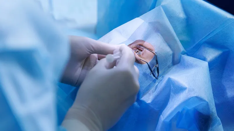
For millions of people, LASIK surgery offers the freedom to see clearly without the need for glasses or contact lenses. This life-changing procedure has revolutionized the treatment of refractive errors like nearsightedness, farsightedness, and astigmatism, dramatically improving quality of life. However, as with any surgery, LASIK does come with its risks. One such risk, though rare, is keratectasia—a serious complication that can occur after LASIK and lead to long-term vision problems if not properly managed.
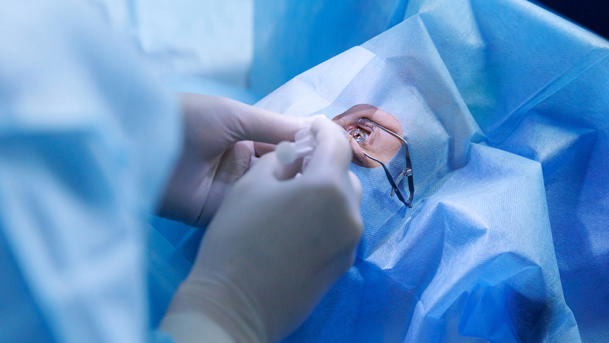
Keratectasia happens when the cornea, the clear front part of the eye, weakens and begins to bulge, often due to excessive tissue removal during LASIK. While keratectasia is uncommon, its impact on vision can be profound, making early detection critical. In this article, we’ll explore what keratectasia is, how it affects vision, the signs to watch out for, and the most effective treatment options. Whether you're considering LASIK or are recovering from the surgery, understanding this condition can help you protect your vision.
What Is Keratectasia? Understanding the Condition
Keratectasia, also known as corneal ectasia, is a condition in which the cornea becomes weakened after LASIK surgery. LASIK works by reshaping the cornea to correct refractive errors, but in some cases, this reshaping can cause the cornea to become too thin. When this occurs, the cornea may begin to bulge outward, distorting vision and leading to symptoms like blurred or distorted eyesight.
While post-LASIK side effects such as dry eyes or mild fluctuations in vision typically resolve on their own, keratectasia is more persistent and can worsen over time without intervention. The condition can be debilitating, especially if left untreated, as it may progress to the point where a corneal transplant is required to restore vision.
Understanding how keratectasia develops and identifying its symptoms early are key to preventing permanent vision loss.
Recognizing the Early Symptoms of Keratectasia
Keratectasia can sometimes be hard to spot right away, as its symptoms may appear gradually. Not all patients experience the same issues, but here are some common signs to be aware of:

Blurry Vision: One of the earliest and most noticeable symptoms, blurry vision can occur even after the LASIK recovery period. If you notice a gradual loss of sharpness, especially in low-light conditions, this could signal keratectasia.
Distorted Vision: Straight lines may appear wavy, or objects could seem out of focus. This distortion happens because the cornea’s shape is no longer uniform.
Increased Sensitivity to Light: As the cornea becomes more irregular, light may scatter, causing glare or halos around lights, especially at night.
Fluctuating Vision: Vision may change throughout the day, with some periods of clear vision followed by blurriness. This fluctuation can be a sign that the cornea is unstable.
Double Vision: In more advanced cases, the distortion in the cornea’s shape can lead to double vision as the light entering the eye is unevenly refracted.
If you’ve undergone LASIK and experience any of these symptoms, it’s essential to consult your ophthalmologist for an evaluation. Early detection using advanced diagnostic tools like corneal topography can identify changes in the cornea’s shape long before the symptoms become severe.
Risk Factors for Keratectasia After LASIK
While keratectasia is rare, several factors can increase the likelihood of its development after LASIK. Understanding these risk factors is crucial for both patients and ophthalmologists to ensure that LASIK is a safe option.
Corneal Thickness: Patients with thinner corneas are more vulnerable to keratectasia. If the cornea is already thin, removing tissue during LASIK may leave it structurally compromised and prone to bulging.
Pre-existing Corneal Conditions: Conditions like keratoconus, where the cornea is abnormally thin and shaped, can significantly increase the risk of keratectasia. Patients with a family history of corneal diseases are also at higher risk.
High Refractive Errors: Individuals with extreme refractive errors (e.g., severe myopia or astigmatism) may require more extensive tissue removal during LASIK, which can weaken the cornea and increase the chances of developing keratectasia.
Age: Younger patients, particularly those under 25, may be at a higher risk, as their corneas are still developing and may not be as stable. However, older patients are not immune to this complication either.
Post-Surgical Factors: Aggressive LASIK treatments or procedures on corneas that are already weakened can contribute to the development of keratectasia. Additionally, insufficient post-operative care and follow-up can hinder early detection.
By carefully assessing these risk factors before surgery, ophthalmologists can minimize the likelihood of complications and ensure the best possible outcome for LASIK patients.
Treatment Options for Keratectasia
The treatment for keratectasia largely depends on how early the condition is detected and the severity of its symptoms. Fortunately, there are several options available that can stabilize the cornea and improve vision.
Corneal Cross-Linking: One of the most promising treatments for keratectasia, corneal cross-linking involves using UV light and riboflavin (vitamin B2) to strengthen the collagen fibers in the cornea. This minimally invasive procedure can halt the progression of the disease and even improve vision in some cases.
Specialized Contact Lenses: For patients with early-stage keratectasia, specialized contact lenses, such as rigid gas-permeable (RGP) lenses or scleral lenses, can help improve vision by creating a smooth, stable surface over the cornea. These lenses are custom-designed to address the unique needs of each patient.
Intacs: In cases where corneal cross-linking alone is not enough, small, curved inserts can be placed in the cornea to flatten the bulging and restore vision. Intacs are often used when keratectasia has caused moderate visual disturbances.
Corneal Transplant: For patients with severe keratectasia that does not respond to other treatments, a corneal transplant may be necessary. During this procedure, the damaged cornea is replaced with a donor cornea, restoring vision but requiring a long recovery period.
Early Detection and Preventive Care
The key to effectively managing keratectasia lies in early detection. The sooner the condition is identified, the more likely it is to be stabilized with less invasive treatments.
In addition to regular post-surgical checkups, patients should choose a clinic that utilizes the latest diagnostic technology, such as corneal topography and pachymetry, to monitor corneal health closely. By detecting changes in the cornea early, ophthalmologists can prevent the progression of keratectasia and help patients maintain clear, stable vision for years to come.
How Jryn Eye Clinic Supports Early Detection
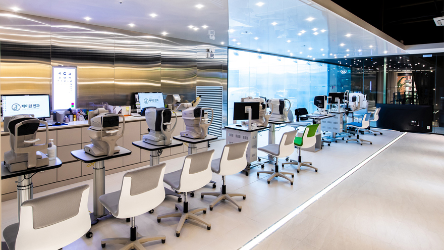
At Jryn Eye Clinic, we are dedicated to providing the highest standard of care to our patients by prioritizing the early detection of complications such as keratectasia after LASIK surgery. Our clinic stands out in the field of ophthalmology, not just because of our advanced technologies, but also due to the expertise of our medical team, which has over 20 years of experience in performing LASIK surgeries and handling their aftercare.
Advanced Diagnostic Tools for Early Detection
One of the primary reasons why our patients trust us for LASIK and post-operative care is our use of cutting-edge diagnostic tools. These tools enable us to track the health of your cornea with precision, detecting even the most subtle changes that might indicate the onset of keratectasia. Our clinic uses:
Corneal Topography: This diagnostic tool provides a detailed map of your cornea, showing the surface’s shape and revealing any irregularities that may suggest the early stages of keratectasia.
Pachymetry: By measuring the thickness of your cornea, pachymetry helps us identify areas of thinning that may signal the development of keratectasia.
Scheimpflug Imaging: This 3D imaging technology offers a detailed view of your corneal layers, enabling us to monitor the structural integrity of your cornea in real-time.
OCT (Optical Coherence Tomography): This advanced imaging system allows us to assess the layers of your cornea in high detail, helping us spot any signs of irregularities or weakening at the earliest possible stage.
These diagnostic technologies provide us with comprehensive insights into your corneal health, allowing us to detect any potential signs of keratectasia well before symptoms manifest.
Expert Monitoring Throughout Your LASIK Journey
Our commitment to the early detection of keratectasia doesn’t stop with the diagnostic tools we use. At Jryn Eye Clinic, we believe in a holistic, patient-focused approach that includes regular follow-up care. Dr. Han Sang Yeop, along with our team of experienced specialists, conducts thorough post-surgical assessments to ensure the cornea is healing properly. For our patients who have undergone LASIK, we emphasize the importance of attending all follow-up appointments so we can monitor your recovery and catch any potential issues early.
Personalized Care for Every Patient
We understand that each patient’s eyes are unique, which is why we offer personalized care that tailors treatments and monitoring schedules to your individual needs. Whether you are considering LASIK or have already undergone the procedure, we take the time to assess your risks and ensure that the treatment plan aligns with your specific circumstances. Our dedicated specialists ensure that you are well-informed and supported throughout your LASIK journey, minimizing the likelihood of complications like keratectasia.
Conclusion: Proactive Steps for Protecting Your Vision
LASIK is a highly effective procedure for correcting refractive errors, but it’s not without its risks. Keratectasia, though rare, is a serious complication that can affect vision if not detected and treated early. The key to preserving vision after LASIK is being proactive about your eye health. Recognize the symptoms, understand the risk factors, and seek immediate attention if you notice any changes in your vision after surgery.
Choosing a clinic with advanced diagnostic tools and experienced surgeons is crucial to ensuring the best possible outcomes. If you're experiencing any symptoms of keratectasia or are considering LASIK, make sure to schedule a consultation with a skilled ophthalmologist who can guide you through the process and ensure your eyes are in good hands.
Ready for a Personalized 1:1 Eye Consultation?
Schedule your comprehensive 3D eye examination with Chief Director Dr. Sang-Youp Han and our ophthalmology team on the 14th floor of JRYN Medical Center in Seomyeon, Busan.