Jryn Eye Clinic Busan (Seomyeon) · SMILE LASIK · EVO+ Visian ICL · Cataract & Retina Center · Tel: +82-51-710-1004
en-US

1:1 Consultation
CLINICAL ARTICLE & GUIDE
Cataract Surgery When You Have Keratoconus: Special Tips
Discover expert cataract surgery tips for keratoconus patients. Learn how to protect vision, choose lenses wisely, and ensure better outcomes. Read more now!

Introduction
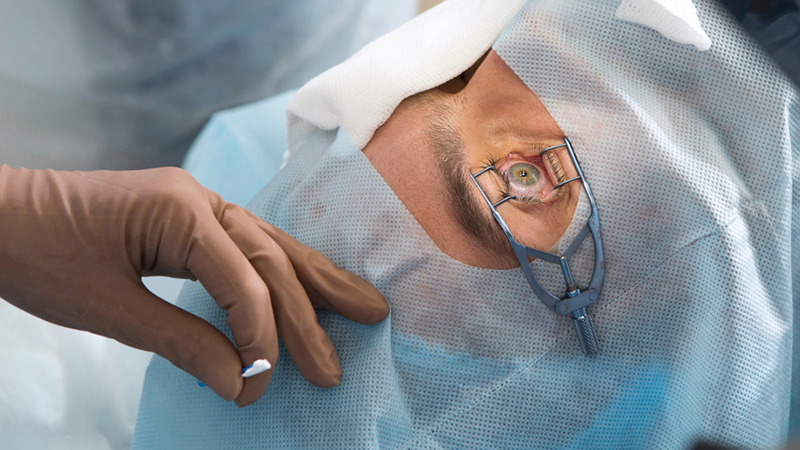
If you're navigating both keratoconus and cataracts, you may be feeling uncertain about your next steps. It's not just about blurry vision anymore—it's about making decisions in a uniquely complex situation. At Jryn Eye Clinic in Busan, we often meet patients in your shoes: people who’ve spent years managing keratoconus, only to now be told they also have cataracts. This isn’t rare, and you’re not alone. But it does call for a more nuanced, precision-driven approach to surgery.
This article walks you through what cataract surgery looks like when keratoconus is part of the picture—what changes, what you need to watch for, and how we approach these cases at our clinic. Our goal? To help you see clearly again—not just physically, but with clarity about your options and what lies ahead.
Understanding the Basics: Cataracts + Keratoconus
Keratoconus causes your cornea—the clear front surface of the eye—to thin and bulge outward in a cone-like shape. This leads to distorted, blurry, and often fluctuating vision. Cataracts, meanwhile, are the clouding of the eye’s natural lens. It’s a condition we all face as we age, typically starting in the 60s and beyond, but sometimes appearing earlier, especially in keratoconus patients who've undergone long-term contact lens wear or eye rubbing.
When both occur in the same eye, cataract surgery becomes more than routine. Why? Because the irregular shape of a keratoconic cornea changes how we measure, plan, and perform the procedure. What seems straightforward in a normal eye becomes a multi-layered clinical puzzle.
Why Cataract Surgery Is More Complex with Keratoconus?
1. Unreliable Biometry and IOL Calculations
Standard cataract surgery relies heavily on accurate measurements of the eye’s dimensions—especially corneal curvature (keratometry). But in keratoconus, these values are often unreliable due to the irregular, steep corneal shape. This can lead to inaccurate predictions for the power of the intraocular lens (IOL), increasing the risk of a post-operative “refractive surprise.” Even a few diopters of error can make a significant difference in visual outcomes.
To make matters more complicated, the posterior curvature of the cornea in keratoconus patients is often disproportionately steep, which is not accounted for in standard formulas. That’s why relying on a single measurement or conventional calculation can be misleading—and why clinics like ours use multiple methods and repeated scans to cross-check every number.
2. Irregular Astigmatism Complicates Vision Correction
Keratoconus induces an astigmatism that is often irregular—not simply a cylindrical blur that glasses can correct, but a wave-like distortion requiring specialty contact lenses. Even after successful cataract removal, some patients may still need rigid gas-permeable (RGP) or scleral lenses for best vision.
We always make sure patients understand that lens implantation won’t “fix” the irregular cornea. If you’re someone who currently relies on RGPs or scleral lenses, you may very well need to return to them post-surgery—though your clarity and contrast sensitivity could still be much improved due to the removal of the cloudy cataract.
3. Structural Fragility of the Cornea
The thinner, weaker cornea poses a risk during surgery. Standard clear-corneal incisions might induce more astigmatism or compromise healing. Adjustments in surgical technique are essential to maintain the structural integrity of the eye. For example, we may opt for a scleral tunnel incision instead of a typical clear corneal one.
Even the intraocular pressure changes during phacoemulsification (the technique used to break up and remove the cataract) can place additional strain on an already-compromised corneal structure. We address this by adjusting fluidics and using more viscoelastic materials to support the corneal dome during surgery.
4. Progression Risk
Keratoconus is progressive in many young patients. If it hasn’t stabilized—or if no prior cross-linking has been done—cataract surgery could be followed by further corneal changes. That’s why we prefer to confirm stability before proceeding. If there's any doubt, we may recommend corneal collagen cross-linking (CXL) first to prevent further progression.
And here's something not often discussed: post-cataract visual shifts can mask ongoing keratoconus progression. Just because the cataract is gone and the vision seems sharper doesn’t mean the cornea has stopped changing. Long-term follow-up is key.
5. Limitations of Premium IOLs
While multifocal or toric IOLs are popular in regular cataract cases, they often perform poorly in keratoconic eyes. Multifocal lenses, in particular, can reduce contrast sensitivity and worsen halos or glare in already compromised corneas. Toric lenses may be helpful in select cases—specifically mild, stable keratoconus with regular astigmatism—but even then, their use must be approached with caution.
This is why we emphasize function over marketing. Premium IOLs may sound attractive, but in complex eyes, simplicity and precision often produce the best outcomes.
How We Handle Cataract Surgery in Keratoconus at Jryn Eye Clinic?
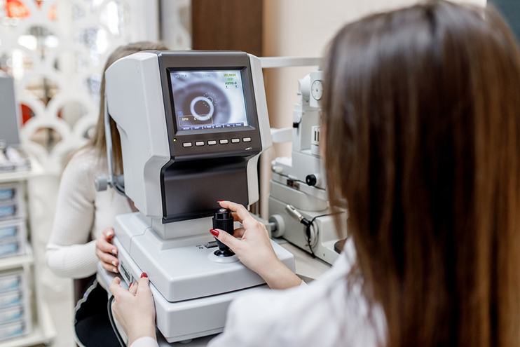
At Jryn Eye Clinic, our process is thorough, evidence-based, and deeply personalized. Here's how we approach these cases:
Step 1: Corneal Stability Check
Before surgery, we analyze serial corneal topography or tomography images over several months. If there’s any indication of progression, corneal cross-linking (CXL) may be recommended before proceeding with cataract surgery. We also check for subclinical signs of steepening that might be missed on a single visit.
Step 2: Enhanced Biometry and Imaging
We use advanced imaging tools like Pentacam and IOLMaster 700 to assess both anterior and posterior corneal curvature. Multiple readings are averaged to reduce error margins. We also apply keratoconus-specific IOL calculation formulas such as Barrett True-K or Kane Keratoconus Formula.
These tools help us avoid common pitfalls, such as overestimating corneal power, which could lead to selecting an IOL that leaves the patient far-sighted. Even a small miscalculation can lead to months of dissatisfaction post-op—something we are committed to preventing.
Step 3: Realistic Target Setting
Instead of aiming for complete spectacle independence, we often target a slight degree of myopia (nearsightedness). This offers better functional vision and leaves room for correction via contact lenses or glasses, if needed. We also factor in the patient’s dominant eye, lifestyle needs, and previous experience with visual aids.
Step 4: Lens Selection
Monofocal IOLs are our go-to recommendation. Toric IOLs may be suitable in mild, stable keratoconus with regular astigmatism. However, multifocal lenses are generally avoided. The goal is functional clarity—not fancy optics that can worsen visual noise. We also involve the patient in this decision through detailed counseling sessions and visual simulations.
Step 5: Customized Surgical Technique
Depending on corneal shape and thickness, we may:
Use a scleral tunnel instead of a corneal incision
Apply corneal sutures for wound stability
Use trypan blue dye for better capsule visibility
Modify fluidics to minimize stress on the cornea
We plan our incision site carefully to minimize induced astigmatism and avoid structurally weak zones. Every move is measured.
Step 6: Post-Operative Lens Fitting and Monitoring
After surgery, we evaluate vision and determine whether the patient needs glasses, soft lenses, or specialty contacts. We also schedule regular topography to ensure the keratoconus remains stable. If further intervention is needed—such as cross-linking or scleral lens fitting—we coordinate this as part of the long-term care plan.
Patients are never discharged with just “we’ll see how it goes.” We create a roadmap for care that stretches months and even years beyond the operating table.
Real Talk: What Patients Need to Know
Perfect uncorrected vision isn’t always possible. Especially in moderate to severe keratoconus, the goal shifts from “glasses-free” to “best functional vision.”
You may still need contact lenses. Especially if your vision was dependent on RGP or scleral lenses before surgery.
Long-term care continues. Cataract surgery doesn’t eliminate the keratoconus—it just treats one part of the problem.
Choose your surgeon wisely. Experience in both corneal disease and cataract surgery is critical. This isn’t a case for a ‘one-size-fits-all’ approach.
Why Jryn Eye Clinic?

Located in Busanjin-gu, Busan, Jryn Eye Clinic is uniquely positioned to handle complex cases like these. Led by Dr. Han Sang Yeop, a board-certified ophthalmologist with over 20 years of surgical experience, we offer:
Precision diagnostics using high-end topography and biometry tools
Expertise in both corneal diseases and lens surgery
Transparent, thorough patient education
Custom-tailored surgical plans with safety and accuracy at the core
For patients with keratoconus facing cataract surgery, we provide not just a treatment—but a strategy for long-term visual stability. We view every eye not just as a case, but as a story—with history, challenges, and future goals.
Conclusion: Clarity Through Customization
If you have keratoconus and now face cataract surgery, don’t settle for a generic plan. Your vision—and your future—deserve a nuanced approach that accounts for every variable your eyes bring to the table.
At Jryn Eye Clinic, we believe in precision over shortcuts, in informed decisions over assumptions. Schedule a consultation today, and let’s talk about your unique eyes, your goals, and how we can restore clarity—not just in your vision, but in your care path forward. You’re not just a patient—you’re a partner in the process. And when it comes to something as important as your sight, that makes all the difference.
Ready for a Personalized 1:1 Eye Consultation?
Schedule your comprehensive 3D eye examination with Chief Director Dr. Sang-Youp Han and our ophthalmology team on the 14th floor of JRYN Medical Center in Seomyeon, Busan.