Jryn Eye Clinic Busan (Seomyeon) · SMILE LASIK · EVO+ Visian ICL · Cataract & Retina Center · Tel: +82-51-710-1004
en-US

1:1 Consultation
CLINICAL ARTICLE & GUIDE
Advanced Eye Diagnostic Technology vs. Basic Exams
Advanced eye diagnostic technology vs a standard eye exam: how Jryn's Tomey CASIA2, OA-2000, and IOLMaster work-up differs, and why the depth matters.
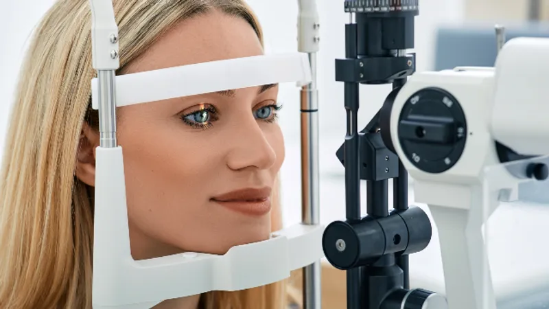
In-Office Diagnostics vs. Standard Eye Exams: Why Jryn's Work-Up Differs
Medically reviewed by Dr. Sang Youp Han, MD, Chief Director, Jryn Eye Clinic · Last updated July 2026
When surgery is on the table, advanced eye diagnostic technology is what separates a routine vision check from a true pre-surgical evaluation. A standard eye exam is designed to answer everyday questions: how well you see and what glasses prescription you need. A surgical work-up has to answer harder ones, such as whether your cornea can safely be reshaped, or exactly what lens power your eye requires. Those decisions rest on precise measurement, not just a reading of the eye chart. This article explains how a standard exam differs from the in-office diagnostic work-up at Jryn Eye Clinic in Busan, South Korea, and why that depth matters before any procedure.
To keep this useful, general information about standard eye exams is drawn from independent sources such as the American Academy of Ophthalmology, while the clinic-specific details come only from what Jryn Eye Clinic documents. No outcomes are promised, and no prices are quoted.
What a Standard Eye Exam Covers
- A standard comprehensive eye exam is a valuable, everyday tool. As the American Academy of Ophthalmology describes, it typically includes visual acuity testing (reading an eye chart), a refraction test using a phoropter to determine your best glasses or contact-lens prescription, a check of how your pupils respond to light, and often dilation so the doctor can examine the retina and optic nerve. It is comfortable, relatively quick, and well suited to its purpose: checking your sight, updating a prescription, and screening for eye disease.
What a standard exam is not primarily designed to do is produce the fine-grained surgical measurements needed to plan a refractive or cataract operation. That is not a shortcoming; it is simply a different job. Planning surgery requires a deeper layer of imaging and biometry, which is where a dedicated pre-surgical work-up comes in.
What a Pre-Surgical Work-Up Adds
Before vision-correction or cataract surgery, the eye has to be mapped and measured in detail. Two categories of information matter especially:
- Corneal and anterior-segment imaging — the shape, thickness, and internal structure of the front of the eye, which influence whether and how the cornea can be treated.
- Biometry — precise measurements of the eye's dimensions, which for cataract surgery determine the power of the intraocular lens (IOL) to implant.
These measurements do two things at once: they confirm whether you are a candidate for a given procedure, and they feed the surgical plan itself. The American Academy of Ophthalmology notes that accurate measurement of the eye is central to selecting the right IOL in cataract surgery, and the US Food and Drug Administration's LASIK program materials likewise emphasize a thorough pre-operative evaluation. A single visit that includes this depth of measurement is what makes it reasonable to plan surgery.
Inside Jryn Eye Clinic's Diagnostic Line-Up
Jryn Eye Clinic names the specific devices in its work-up, which is what allows the depth to be verified rather than just described. Three are central:
- Tomey CASIA2 — an anterior-segment OCT (optical coherence tomography) system that captures a series of high-speed scans to reconstruct the eye's anterior chamber in 3D and 360 degrees, from the corneal surface to the back of the lens. The clinic uses this imaging for both cataract and lens-implantation (ICL) planning, and states it was the first in Korea, and first in Busan, to introduce the system.
- Tomey OA-2000 — a biometer the clinic describes as measuring corneal thickness, refractive power, anterior chamber depth, lens opacity, and axial length in a single pass, using a wavelength chosen for tissue penetration. The clinic references a Tomey CASIA2 by OA-2000 cross-connection setup used in pre-operative measurement.
- ZEISS IOLMaster 500 — a non-contact optical biometer used for IOL power calculation. The clinic states this optical approach is substantially more accurate than older ultrasound-based measurement, including in denser cataracts, and that it uses an established calculation formula suite.
The clinic also references a visual-quality analyzer used to assess the quality of vision objectively as part of planning. Taken together, this is a measurement stack aimed at candidacy and surgical planning, not just a sight check.
A Side-by-Side View
Because the two kinds of visit are easy to conflate, it helps to see them next to each other. The comparison below is about purpose and depth, not about one being better than the other; each is well suited to its own job.
| Standard eye exam | Pre-surgical work-up at Jryn | |
|---|---|---|
| Main purpose | Check sight, update prescription, screen for disease | Confirm surgical candidacy and plan the operation |
| Typical measurements | Visual acuity, refraction, pupil response, dilation | Detailed anterior-segment imaging plus precise biometry |
| Corneal detail | General assessment | 3D anterior-segment imaging (Tomey CASIA2) |
| Eye dimensions | Not the primary focus | Axial length, anterior chamber depth, corneal thickness (Tomey OA-2000) |
| Lens power planning | Not applicable | Non-contact optical biometry for IOL calculation (ZEISS IOLMaster 500) |
| Typical length | Relatively short | Roughly two hours for the refractive work-up |
The right takeaway is not that a standard exam is inadequate; it is that surgery asks a different question and therefore needs a different depth of measurement.
How the Devices Work Together
No single instrument answers every question, which is why a surgical work-up combines several. The anterior-segment OCT builds a detailed three-dimensional picture of the front of the eye, the biometer supplies the precise dimensions, and the optical biometer focuses on the lens-power calculation that cataract and lens surgery depend on. Jryn Eye Clinic references a cross-connection between its Tomey CASIA2 and OA-2000 systems in pre-operative measurement, meaning the imaging and biometry are used together rather than in isolation. The clinic also references an objective visual-quality analyzer as part of its planning, so that the assessment considers not only the numbers but the quality of vision itself.
What this combination produces is a fuller portrait of the eye than any one test could give. For a refractive patient, that portrait shows whether the cornea can be safely treated and by which technique. For a cataract or lens patient, it drives an accurate lens choice. And for someone considering an implantable collamer lens, the detailed picture of the space inside the front of the eye is directly relevant. The value is in the integration, not in any single gadget.
Why This Depth Matters Before Surgery
The reason to care about all this is practical. Refractive surgery reshapes the cornea, so knowing corneal thickness and structure in detail is what makes it safe to proceed, or a reason to choose a different technique. Cataract and lens surgery replace the eye's lens, so the accuracy of the biometry directly affects how well the implanted lens matches your eye. Detailed anterior-segment imaging also matters for planning an implantable collamer lens, where the space inside the front of the eye is a genuine consideration.
In short, the quality of the measurement shapes the quality of the plan. A very fast, surface-level assessment might be fine for updating glasses, but it does not carry the same information a surgeon needs to plan an operation. That is the core distinction between a standard exam and a surgical work-up.
Diagnostics Also Decide Candidacy
There is a second, equally important role for this depth: it determines whether a given procedure fits you at all. Because Jryn Eye Clinic offers several vision-correction technologies (SMILE, LASIK, LASEK, and EVO Visian ICL) and several cataract approaches, the diagnostic findings are what point toward the appropriate option rather than a one-size choice. A thorough work-up can reveal, for example, that a corneal-laser approach is not ideal and that an implantable lens deserves discussion instead. This is why candidacy is decided from current measurements, not from assumptions or a past summary. You can review the clinic's range of procedures on the Jryn Eye Clinic website.
What to Expect at the Visit
A pre-surgical evaluation is more involved than a routine exam, and a little preparation helps. For its laser vision-correction work-up, the clinic asks patients to stop wearing soft contact lenses for about a week beforehand, and hard or gas-permeable lenses for about two weeks, so the cornea returns to its natural shape for accurate measurement. The clinic describes its refractive pre-operative exam as taking roughly two hours, with same-day results used to discuss whether surgery is advisable. Planning for a longer, more detailed visit than a standard eye check is the right expectation.
For international patients, this evaluation is a distinct, in-person step, and for its vision-correction procedures the clinic generally advises staying in Korea for 3 to 7 days to allow for the procedure and initial follow-up. To arrange an evaluation or ask what to prepare, use the clinic's online inquiry and contact page.
The Bottom Line
- A standard eye exam checks your sight and prescription; a surgical work-up adds detailed imaging and biometry to plan an operation.
- Jryn Eye Clinic names its diagnostic devices, including a Tomey CASIA2 anterior-segment OCT, a Tomey OA-2000 biometer, and a ZEISS IOLMaster 500.
- Precise corneal imaging and biometry make surgery safer to plan and help match the right lens power or procedure to your eye.
- The same measurements determine candidacy, pointing toward the appropriate option among several the clinic offers.
- Expect a longer, more detailed visit than a routine exam, with contact-lens-free preparation beforehand.
Frequently Asked Questions
**Isn't a regular eye exam enough before surgery?**A standard exam is excellent for checking sight and updating a prescription, but it is not primarily designed to produce the detailed corneal imaging and biometry needed to plan surgery. A pre-surgical work-up adds that depth, which is what candidacy decisions and surgical planning rely on.
**What does the Tomey CASIA2 actually do?**It is an anterior-segment OCT that captures high-speed scans to build a 3D, 360-degree picture of the front of the eye, from the corneal surface to the back of the lens. Jryn Eye Clinic uses this imaging for both cataract and ICL planning and states it was the first in Korea to introduce the system.
**Why is biometry so important for cataract surgery?**Cataract surgery replaces the eye's natural lens with an intraocular lens, and the power of that lens must match your eye. Accurate biometry, such as that from the Tomey OA-2000 and ZEISS IOLMaster 500, is central to choosing the right lens, which is why optical measurement is emphasized over older methods.
**Do I need to stop wearing contact lenses before the exam?**Yes, for the laser vision-correction work-up the clinic asks patients to stop soft lenses for about a week and hard or gas-permeable lenses for about two weeks beforehand. Contacts temporarily change the corneal shape, so removing them in advance allows accurate measurement.
**How long does the pre-surgical exam take?**The clinic describes its refractive pre-operative exam as taking roughly two hours, with same-day results used to discuss whether surgery is advisable. That is considerably longer than a routine eye exam, reflecting the added imaging and measurement involved.
**Can the diagnostics tell me which procedure I need?**The findings guide that decision. Because the clinic offers several vision-correction and cataract options, the measurements help point toward the procedure that fits your eyes, rather than forcing one choice. The final recommendation is made with the surgeon based on your results.
This article is for general informational purposes and does not constitute individualized medical advice. Candidacy, technique, and expected outcomes can only be determined through a consultation with a qualified eye surgeon.
Ready for a Personalized 1:1 Eye Consultation?
Schedule your comprehensive 3D eye examination with Chief Director Dr. Sang-Youp Han and our ophthalmology team on the 14th floor of JRYN Medical Center in Seomyeon, Busan.